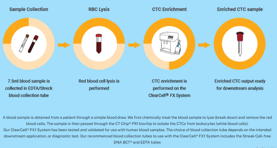
A blood sample is obtained from a patient through a simple blood draw. We first chemically treat the blood sample to lyse (break down) and remove the red blood cells. The sample is then passed through the CT Chip® FR1 biochip to isolate the CTCs from leukocytes (white blood cells).
Our ClearCell® FX1 System has been tested and validated for use with human blood samples. The choice of blood collection tube depends on the intended downstream application, or diagnostic test. Our recommenced blood collection tubes to use with the ClearCell® FX1 System includes the Streak Cell-free DNA BCT® and EDTA tubes.
1.Label-Free enrichment
Recovers heterogeneous populations of CTCs
Ready integration to various downstream applications
2. Continuous sample flow
Intact and viable cells
3. Automated system
Consistent and reproducible
Fast processing time
A blood sample is obtained from a patient through a simple blood draw. We first chemically treat the blood sample to lyse (break down) and remove the red blood cells. The sample is then passed through the CT Chip® FR1 biochip to isolate the CTCs from leukocytes (white blood cells). Our ClearCell® FX1 System has been tested and validated for use with human blood samples. The choice of blood collection tube depends on the intended downstream application, or diagnostic test. Our recommenced blood collection tubes to use with the ClearCell® FX1 System includes the Streak Cell-free DNA BCT® and EDTA tubes
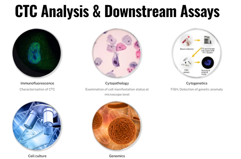
Fluorescently-labelled antibodies specific to CTCs and leukocytes can be used to identify and enumerate CTCs or detect cancer-specific biomarkers. CTC counts provide prognostic information, particularly, the metastatic aggressiveness of the tumour. An increase in CTC counts may suggest tumour relapse after therapeutic intervention.
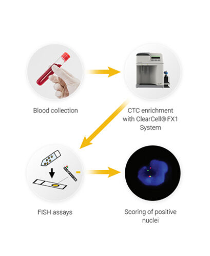
CTCs isolated using our ClearCell® FX1 System can be analysed using fluorescent in situ hybridisation (“FISH”), a technique that allows pathologists to identify chromosomal abnormalities in cells, including gene translocation, amplification and deletions. In a FISH assay, fluorescent probes (labelled fragments of DNA or RNA) that bind to specific nucleotide sequences in chromosomes are applied to the CTCs. The presence or absence of specific nucleotide sequences is detected by noting the location and counts of fluorescent dots after the probes have been applied. FISH can be used to detect ALK gene rearrangement in lung cancer patients and HER2 gene amplification in breast cancer patients, in order to help physicians, identify patients that are suitable for ALK and HER2 targeted drug therapies (for example, the drug Crizotinib targets ALK in lung cancer patients and the drug Trastuzumab targets HER2 in breast cancer patients).
Gene mutations and aberrant gene expression are commonly found in cancer cells. Some cancer mutations are drug-actionable targets that can be used to select effective treatment strategies. An example would be the detection of BRAF V600E mutation in melanoma (skin cancer) patients, which can be targeted by the drug Vemurafenib.